Newsletter Subscribe
Enter your email address below and subscribe to our newsletter
Enter your email address below and subscribe to our newsletter

Discover the key insights on mandibular tori removal, including when it's necessary, the surgical process, recovery tips, and how to manage potential complications. Perfect for patients seeking relief and a smoother dental experience.
Mandibular tori, those bony growths that can appear along the inner surface of the lower jaw, often go unnoticed until they start to interfere with daily life.
Whether it’s making dental procedures more complicated, causing discomfort, or affecting the fit of dentures, the decision to remove these benign but bothersome protrusions can significantly improve a patient’s quality of life.
In this comprehensive guide, we delve into the intricacies of mandibular tori removal, offering essential insights into when removal is advised, what the surgical process entails, and how to navigate the recovery phase effectively.
We also provide practical advice on managing any potential complications, ensuring you’re fully informed and prepared for a smoother dental experience.
If you’re contemplating surgery or simply curious about your options, this article is your go-to resource for understanding mandibular tori removal.
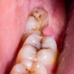
Mandibular tori (torus mandibularis, if it’s one) are bony growths located on the inner side of the lower jaw, below and near the tongue. They are generally benign and vary in size.
Overgrowth of mandibular tori can lead to several symptoms or issues, including:
Mandibular tori removal is not always necessary, as these bony growths are generally benign. However, there are specific circumstances under which removal might be considered or required:
If the tori cause significant discomfort or pain during eating, speaking, or other activities, removal might be recommended to alleviate these symptoms.
Large tori can interfere with chewing, speaking, and the overall comfort of the mouth. If they significantly affect oral functions, removal can help restore normal function and comfort.
If the presence of tori makes it difficult to maintain good oral hygiene, leading to recurrent oral health issues like plaque buildup, tooth decay, or periodontal disease, removal might be considered to facilitate better oral care.
Mandibular tori can interfere with the fitting and comfort of dentures or other dental appliances. If tori prevent proper fitting or cause discomfort with dentures, removal might be necessary for a better fit and function.
Although rare, if the tori are subject to frequent trauma leading to ulceration or infection of the overlying mucosa, removal might be recommended to prevent further complications.
In some cases, if the tori are large and cause significant aesthetic concerns for the patient, removal might be considered for cosmetic reasons.
Benefits of mandibular tori removal
Mandibular tori removal can be performed using different surgical techniques, depending on the size and location of the tori, the patient’s overall health, and the surgeon’s preference and expertise.
Here are the primary types or techniques of mandibular tori removal:
This method involves making an incision over the tori, exposing the bone, and then using surgical instruments to remove the bony growth. The area is smoothed, and the incision is sutured closed.
Suitable for tori of various sizes, especially larger ones that require significant bone removal.
Laser surgery uses a focused beam of light to cut tissue and remove the tori. It can seal blood vessels, reducing bleeding during the procedure.
Often used for smaller tori or in situations where precision and minimal invasiveness are desired. It’s noted for reduced postoperative discomfort and faster healing times.
Tori removal surgery, also known as torus mandibularis removal or excision, is a procedure performed to remove bony growths in the mouth, specifically on the inner side of the lower jaw (mandibular tori) or the roof of the mouth (palatal tori).
The surgery aims to alleviate discomfort, improve oral function, or prepare the mouth for dental prostheses.
Here’s a general overview of how the surgery is performed:
The procedure is typically performed under local anesthesia, which numbs the area around the tori. In some cases, sedation or general anesthesia may be used, especially if the patient is anxious or if the tori are large.
Recovery from mandibular tori removal varies among individuals, depending on the extent of the surgery, the individual’s overall health, and the specific technique used for removal.
Here are some general guidelines and expectations for the recovery period:
While complications are rare, it’s important to be aware of signs that may indicate a problem:
The cost of oral tori removal can vary widely depending on several factors, including the geographic location of the dental practice or oral surgeon’s office, the complexity of the case, the specific technique used for the removal, the anesthesia type, and whether additional treatments are required in conjunction with the tori removal.
The cost for oral tori removal can range from $600 to several thousand dollars. The wide range reflects differences in surgical approaches (e.g., traditional surgery vs. laser surgery), the number of tori being removed, and the need for any specialized care.
Many dental insurance plans consider tori removal to be a covered procedure, especially if it is deemed medically necessary rather than purely cosmetic. For example, if the tori interfere with oral functions, cause pain, or prevent the proper fitting of dentures, insurance may cover part or all of the procedure.
In some cases, if there’s a medical reason for the removal, medical insurance might cover the procedure. This is less common and depends on the specifics of the insurance policy.
It’s often necessary to get pre-authorization from the insurance company before the procedure. The dental office or surgeon’s office may assist with this process, providing necessary documentation to demonstrate the medical necessity of the procedure.
FIND OUT MORE: How To Get Dental Insurance without a Job
Mandibular tori removal pain varies individually but is generally manageable with local anesthesia during the procedure, minimizing discomfort. Postoperative pain can be expected but is typically controlled with prescribed pain relievers or over-the-counter medications. Most patients report the pain as mild to moderate, subsiding significantly within the first few days after surgery. Proper postoperative care can further alleviate discomfort and promote healing.
Recovery from tori surgery typically takes a few weeks for initial healing, with complete bone healing possibly taking several months. Most patients can resume normal activities within a few days, although dietary restrictions and oral hygiene precautions are advised to support healing.
Deciding to remove tori depends on whether they cause discomfort, interfere with oral functions, or affect dental appliance fitting. If tori lead to pain, difficulty eating, or oral hygiene challenges, removal might be considered. Consult with a dental professional to evaluate your specific situation and discuss potential benefits and risks.
There’s no proven method to stop the growth of mandibular tori, as their development is influenced by factors like genetics, jaw structure, and possibly stress on the jawbones. Maintaining good oral hygiene and regular dental check-ups can help monitor their size and address any issues early.
After torus removal, it’s uncommon but possible for the torus to regrow, especially if underlying factors like genetic predisposition or specific jaw stressors persist. Regular dental check-ups can help monitor any changes and address potential regrowth early.
Fact Checked
Our dedicated team rigorously evaluates every article and guide to ensure the information is factual, up-to-date, and free of bias.
Updated Regularly
We update our articles and reviews regularly to ensure you have access to the latest data in the dental industry.
The content on Dental3DU’s blog is intended for educational purposes only. This information should not be relied upon as professional medical counsel. Be sure to always consult with your dentist about the dangers and benefits of any medication, treatment or procedure.
Dental articles in your inbox. Subscribe